Recommend Posts

Science
Causal Inference Engine: Debunking the Disease Attribution Model Based on Correlation
By /Jul 18, 2025
For decades, medical research has relied heavily on statistical
correlations to identify potential risk factors for diseases.
Observational studies linking smoking to lung cancer or
cholesterol to heart disease have undoubtedly saved millions of
lives. However, as we venture into the era of precision
medicine, the limitations of correlation-based approaches are
becoming increasingly apparent. The emergence of causal
inference engines promises to revolutionize how we understand
disease etiology by distinguishing true causation from mere
association.

Science
Re-Analysis of Obsolete Experimental Records with AI
By /Jul 18, 2025
In the quiet corners of research laboratories and university
archives, countless volumes of handwritten experimental logs and
typewritten reports gather dust. These forgotten records, often
spanning decades, contain a wealth of untapped scientific data
that could hold the key to breakthroughs in fields ranging from
medicine to materials science. What if artificial intelligence
could breathe new life into these neglected archives?

Science
Compound Eye Camera: A Panoramic Imaging Device Inspired by Insect Vision
By /Jul 18, 2025
In the quest to develop advanced imaging systems, scientists and
engineers are increasingly turning to nature for inspiration.
One of the most fascinating breakthroughs in this field is the
development of compound eye cameras, which mimic the visual
systems of insects. These cameras, inspired by the intricate
structure of insect eyes, promise to revolutionize panoramic
imaging with their wide field of view, exceptional motion
detection, and compact design.
Science
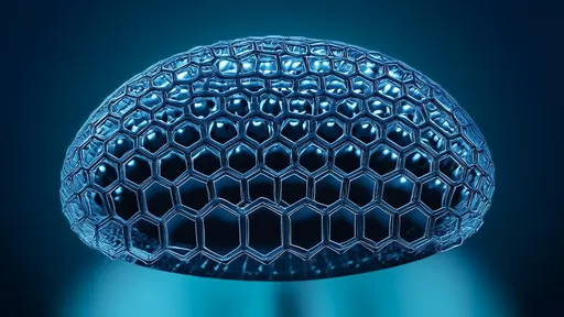
BEETLE-INSPIRED WATER-COLLECTING AIR CONDENSER WITH IMITATIVE NANOSTRUCTURES
By /Jul 18, 2025
In the realm of biomimicry, few innovations have captured the
imagination of scientists and engineers quite like the
water-harvesting techniques of the Namib Desert beetle. This
unassuming insect, thriving in one of the planet's most arid
regions, has inspired a groundbreaking approach to atmospheric
water collection. Researchers are now translating the beetle's
unique surface morphology into advanced nano-engineered
materials, paving the way for a new generation of air condensers
that could revolutionize water scarcity solutions.
Science
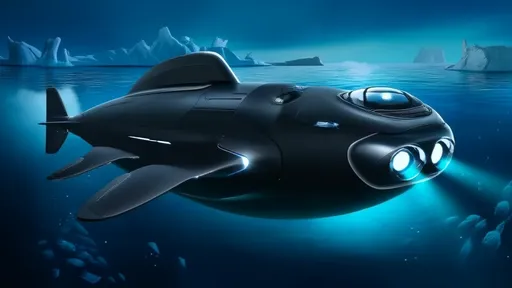
Penguin Submarine: Bionic Vortex Ring Propulsion for Polar Explorers
By /Jul 18, 2025
The frigid, uncharted depths of Earth's polar regions have long
posed a challenge for scientists and explorers. Traditional
underwater vehicles, while effective in temperate waters, often
struggle with the extreme conditions found beneath Arctic and
Antarctic ice. But inspiration has emerged from an unlikely
source: the humble penguin. Engineers and marine biologists have
collaborated to develop the Penguin Submarine, a revolutionary
polar explorer that mimics the efficient propulsion methods of
its avian namesake.
Science
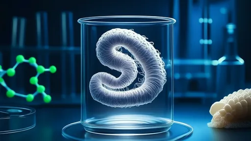
Spider Silk Artificial Tendons: Biomaterials from Transgenic Silkworm Protein
By /Jul 18, 2025
In a groundbreaking development at the intersection of
biotechnology and materials science, researchers have
successfully engineered transgenic silkworms to produce spider
silk proteins. This innovation opens new frontiers in creating
artificial tendons with unprecedented strength and elasticity.
The fusion of centuries-old sericulture with cutting-edge
genetic engineering may soon revolutionize medical implants and
high-performance textiles.

Science
Shark Skin Hull: Micro-Ribbed Drag Reduction for Fuel Saving
By /Jul 18, 2025
The shipping industry has long been plagued by the enormous fuel
costs associated with vessel operation. With global trade
relying heavily on maritime transport, even minor improvements
in fuel efficiency can translate into significant financial and
environmental benefits. One of the most promising innovations in
this field is the application of shark-inspired micro-riblet
coatings on cargo ships—a technology that reduces hydrodynamic
drag and slashes fuel consumption.

Science
Regional Climate Simulation: Century-Long Forecasts on a Square Kilometer Grid
By /Jul 18, 2025
The scientific community has reached a pivotal moment in climate
modeling, with researchers now capable of simulating regional
climate patterns at an unprecedented resolution of one square
kilometer over century-long timescales. This breakthrough
represents a quantum leap from traditional models that operated
at coarser resolutions, often missing critical local-scale
phenomena that drive weather extremes and long-term climatic
shifts.

Science
Mangrove Gene Editing: Cross-Species Delivery of Salt-Tolerance Genes
By /Jul 18, 2025
The scientific community is abuzz with groundbreaking research
exploring the potential of cross-species gene transfer,
particularly focusing on salt-tolerant genes from mangrove
ecosystems. This cutting-edge approach could revolutionize
agriculture in saline-affected regions worldwide, offering hope
for food security in the face of climate change-induced soil
salinization.
Science
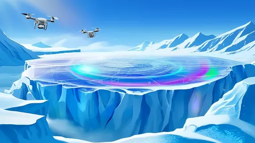
Glacial Protection Coating: Nano Reflective Material for Ice Melting Inhibition
By /Jul 18, 2025
The accelerating retreat of glaciers worldwide has spurred
scientific innovation in cryospheric preservation technologies.
Among the most promising developments is the emergence of
nano-reflective coatings designed to mitigate ice melt through
advanced photonic engineering. These sophisticated materials
represent a convergence of materials science, climatology, and
nanotechnology, offering a potential tool for slowing glacial
mass loss in vulnerable regions.
Science
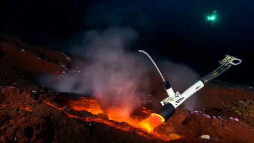
Basalt Carbonation: Geological Reactor for CO₂ Sequestration
By /Jul 18, 2025
In the quest to combat climate change, scientists and engineers
are turning to the Earth itself for solutions. One of the most
promising avenues is basalt carbon mineralization, a natural
process accelerated to trap carbon dioxide (CO₂) permanently in
rock. This method leverages the chemical reactivity of basaltic
rocks, which are abundant worldwide, to convert CO₂ into stable
carbonate minerals. The concept is simple yet profound: mimic
and enhance Earth’s own carbon sequestration mechanisms to
address human-induced emissions.

Science
Marine Whitening Project: Aerosol Enhancement of Cloud Albedo
By /Jul 18, 2025
The concept of brightening Earth’s clouds to reflect more
sunlight back into space is no longer confined to the realm of
speculative science fiction. Known as marine cloud brightening
(MCB), this geoengineering approach has gained traction among
researchers as a potential tool to mitigate global warming. By
injecting sea salt aerosols into low-lying marine clouds,
scientists aim to enhance their albedo—effectively turning them
into larger, more reflective mirrors that could offset some of
the planet’s rising temperatures. While the idea is
theoretically sound, its real-world implications, ethical
considerations, and technical challenges remain hotly debated.
Science
Transparent Cranial Window: A Living Platform for Long-Term Observation of Gliomas
By /Jul 18, 2025
Glioblastoma and other aggressive brain tumors have long posed
significant challenges for researchers and clinicians. The
complexity of these malignancies, coupled with the difficulty of
monitoring their progression in real time, has hindered the
development of effective treatments. However, a groundbreaking
innovation—the transparent skull window—is transforming the
landscape of glioma research by enabling scientists to observe
tumor dynamics in living subjects over extended periods.
Science
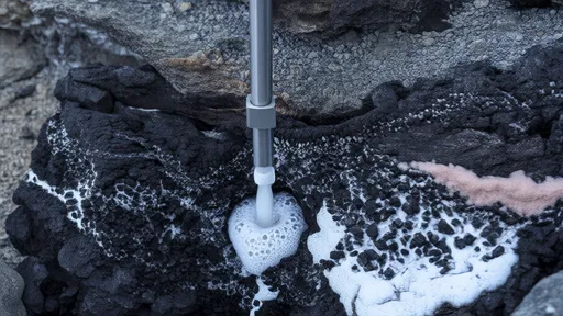
In-situ Analysis of Chemical Gradients at Deep-sea Hydrothermal Vents
By /Jul 18, 2025
The deep ocean remains one of Earth's most enigmatic frontiers,
with hydrothermal vents serving as dynamic hotspots of chemical
and biological activity. Recent advancements in deep-sea mass
spectrometry have enabled scientists to conduct in situ analyses
of these extreme environments, revealing intricate chemical
gradients that were previously inaccessible. The development of
the Deep-Sea Mass Spectrometry Nest (DSMS Nest) represents a
groundbreaking leap in marine science, allowing researchers to
capture real-time data without the need for sample retrieval.

Science
Neutron Holography: Penetrating Non-destructive Imaging of the Qin Shi Huang Mausoleum
By /Jul 18, 2025
The sealed mausoleum of Qin Shi Huang, China's legendary first
emperor who unified the warring states in 221 BCE, has remained
one of archaeology's most tantalizing enigmas. For over two
millennia, the underground complex guarded by the Terracotta
Army has resisted conventional excavation due to both
preservation concerns and the Chinese government's cautious
approach. Now, an unprecedented scientific collaboration is
deploying neutron holography - a cutting-edge imaging technique
- to virtually unveil the tomb's secrets without disturbing a
single artifact.

Science
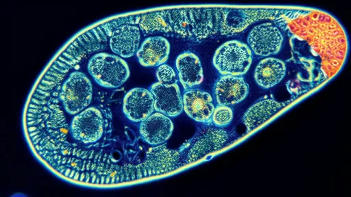
Cryo-Electron Microscopy Cloud: A Globally Shared Protein Structure Library
By /Jul 18, 2025
The world of structural biology has undergone a quiet revolution
in the past decade, with cryo-electron microscopy (cryo-EM)
emerging as the powerhouse technique for visualizing
biomolecules at near-atomic resolution. This technological leap
has coincided with the rise of a remarkable global resource –
the Protein Data Bank (PDB) – which has evolved into a living
atlas of three-dimensional protein structures freely available
to researchers worldwide. The intersection of these two
phenomena is reshaping how we understand life's molecular
machinery.

Science
Attosecond Laser Knife: Precision Surgery for Selective Molecular Bonds
By /Jul 18, 2025
In the realm of ultrafast science, a revolutionary tool has
emerged with the potential to redefine precision at the
molecular level. The attosecond laser scalpel, often referred to
as the "light scalpel," operates on timescales so brief they
defy conventional intuition. Unlike traditional surgical tools
or even femtosecond lasers, this technology targets chemical
bonds with unprecedented selectivity, offering scientists the
ability to perform what can only be described as molecular-scale
surgery.
Science
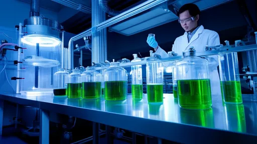
Algae Protein Factory: Super Nutrient Source from Wastewater Cultivation
By /Jul 18, 2025
The concept of turning wastewater into a valuable resource has
long been a dream of environmental scientists and sustainability
advocates. Now, with the emergence of algae-based protein
factories, that vision is becoming a reality. These innovative
facilities are harnessing the nutrient-rich properties of
wastewater to cultivate algae, transforming what was once
considered a waste product into a highly nutritious and
sustainable protein source.